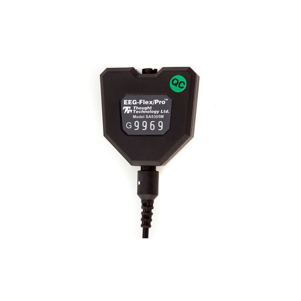
EEG Sensor for use with Thought Technology Procomp Infiniti, Procomp+ or Procomp 2.
See all our our USED products here.
Operating principle
The EEG sensor detects and amplifies the small electrical voltages
that are generated by the brain cells (neurons) when they fire.
Similarly to muscle fibers, neurons of different locations can fire at different rates. The frequencies most commonly looked at, for EEG,
are between 1 and 40 Hz. The EEG sensor records a raw EEG
signal, which is the constantly varying difference of potential
between the positive and negative electrode, and the software
processes that signal by applying a variety of digital filters to the
recorded signal, in order to extract frequency-domain information.
Note: EEG practitioners call the negative electrode "reference" and
the positive electrode, "signal"
Measurement unit
The EEG-Flex/Pro sensor measures micro-Volts (V), raw. A normal EEG signal, recorded from the scalp, will have an amplitude between 0.1 and about 200 V. The raw EEG signal is mainly used to evaluate the quality of the recorded signal and to do artifact rejection. For biofeedback purposes, clinicians will usually want to filter out subsets of the whole EEG bandwidth and measure the real-time changes in the total power generated by all the frequencies in the subset. Generally, the most frequently observed frequency subsets are defined as: High Beta 20- 40 Hz, Beta 15-20 Hz, Sensorimotor rhythm (SMR) 13-15 Hz, Alpha 8-13 Hz, Theta 4-8 Hz and Delta 2-4 Hz.
The frequencies above 40 Hz are interpreted as EMG noise from neighboring muscles.
Sensor placement
The EEG sensor, like EMG and EKG, has three electrodes. The positive one is called the "signal" electrode. The negative one is the reference and the third one is the ground. For EEG applications, the regular Triode, Single Strip or Uni-Gel electrodes are not appropriate.
Prior to placing EEG electrodes on a client's head, it is recommended to gently rub the placement areas, and ear lobes, with a bit of abrasive paste. This will remove the surface layer of dead skin and improve the electrode's contact with the skin. To place the EEG electrodes, fill the tin cups with a generous amount of EEG conductive paste and press it down over the desired spot until the cup is firmly in place. When you do this, some of the paste will be squeezed out of the small hole in the cup; that is expected. You may want to help hold the cup down by pressing on it with a small cotton ball. The cotton ball can be left on the client's head until you remove the electrodes. The monopolar extender cable has one cup and two ear clips. The cup should be placed on an active EEG site - all standard sites for EEG are defined by the international 10-20 EEG electrode placement system (see below). The reference ear clip should be placed on the ear lobe, on the same side as the active site, and the ground ear clip on the ear lobe of the other side. The monopolar placement, frequently called referential placement, is the most commonly used for EEG biofeedback.
A bipolar placement involves recording one signal between two active sites using one sensor. For this placement, you need to place the positive and reference electrodes on two standard EEG locations, usually on the same side of the brain. The ground electrode has an ear clip and should be placed on the earlobe of the opposite side. (Note: bipolar is not the same as bilateral. A bipolar
placement provides only one signal. A bilateral placement
requires two sensors and gives two signals.)
When you want to use two EEG sensors, the electrode placements are similar, except that you do not need to place both ground electrodes
on the client; you can leave one ground lead loose. To get a full understanding of the standard international 10-20 EEG electrode placement system, please refer to professional publications on EEG.
1 Year Warranty
Also available EEG-Z Impedence and EEG Sensor.


